



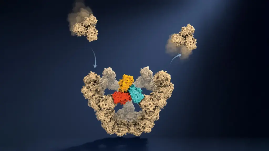
Researchers at the University of Oxford have achieved a groundbreaking milestone in molecular biology by observing virus-like particles assembling themselves one molecule at a time. This unprecedented view has uncovered the surprisingly simple yet elegant rules that allow complex biological structures to form with remarkable reliability. The findings, published in the prestigious journal Nature, offer a deeply detailed look at a dynamic natural process that scientists have struggled to observe directly for decades.
Understanding how viruses construct their protective protein shells—and discovering how that crucial construction phase might be intentionally interrupted—could pave the way for a new generation of antiviral drugs, improved vaccines, and advanced medical treatments. The new discovery helps solve a long-standing biological puzzle, explaining how individual proteins consistently organize themselves into the correct structural geometries despite having thousands of possible ways to connect during the chaotic environment of cellular fluids.
The research team found that viral assembly closely resembles navigating a remarkably complicated maze, where only certain specific pathways lead to lasting progress. In the initial phases of the process, individual protein components form weak connections that can easily break apart if a particular arrangement turns out to be unsuccessful. This inherent structural flexibility allows the molecules to keep trying different combinations continuously until they stumble upon and form particular closed structures. Once multiple stable connections hold these growing structures together, they become far more robust and resistant to falling apart.
These stable arrangements serve as crucial molecular checkpoints, effectively narrowing down the enormous, overwhelming number of possible assembly routes into a much smaller, manageable set of successful pathways. One especially critical milestone occurs when five larger protein building blocks connect together to form a closed pentagonal ring. This configuration represents the first highly stable structure in the overall assembly process. Once this pentagonal cornerstone forms, each subsequent stable stage requires fewer additional building blocks, allowing the viral particle to grow exponentially faster toward completion.
Dr. Roi Asor, co-lead author of the study from the Department of Chemistry and the Kavli Institute for Nanoscience Discovery at the University of Oxford, explained the magnitude of the challenge. "A virus has to solve an extraordinary construction problem," Dr. Asor noted. "Its components somehow have to find the right arrangement among a huge number of possibilities, without a blueprint or machinery directing the process. We can now watch what happens molecule by molecule and see the physical rules that make it possible."
Watching Virus Particles Assemble Molecule by Molecule
To understand the scale of the achievement, it is necessary to examine how viruses naturally store and protect their genetic material. Viruses rely on organized protein shells known as capsids. Depending on the specific type of virus, these resilient protective shells may consist of tens, hundreds, or even thousands of individual protein components working in unison. Remarkably, in many biological cases, these proteins possess the intrinsic capability to arrange themselves into the correct, highly symmetrical structure without any external cellular machinery directing their assembly.
Although scientists have investigated viral self-assembly for decades, capturing the microscopic process as it actively unfolds has presented a major experimental challenge. Many of the vital intermediate structures exist only briefly and appear in extremely small quantities at any given moment. Consequently, traditional scientific methods have generally allowed researchers to examine only the static starting materials and the finished particles, without clearly showing the dynamic transition of how one transforms into the other.
To overcome this persistent limitation, the Oxford researchers studied an engineered virus-like particle composed precisely of 60 individual protein units. They accomplished this by utilizing mass photometry, a cutting-edge analytical technique originally developed at Oxford that determines the precise mass of individual molecules by meticulously measuring the light they scatter. By combining this innovative technology with a novel approach for keeping individual molecules confined and continuously observable, the research team could repeatedly measure the mass of a growing particle in real time as additional proteins attached to the structure.
This sophisticated approach allowed the scientists to follow the construction process precisely as it happened, effectively tracking the continuous addition of individual molecular building blocks over time.
Dan Loewenthal, a co-first author and PhD student in the Department of Chemistry and the Kavli Institute for Nanoscience Discovery at the University of Oxford, emphasized the simplicity and power of the breakthrough, noting that the new method lets them study the assembly process directly by simply taking a video of the molecules in action.
Professor Philipp Kukura, co-lead author of the study from the Department of Chemistry and the Kavli Institute for Nanoscience Discovery at Oxford University, elaborated on how this shifts scientific understanding. "Until now, much of our understanding of how these structures assemble has had to be reconstructed from snapshots or theoretical models," Professor Kukura said. "Being able to both quantify the underlying interactions and follow one particle as it grows changes that. We can see the important intermediate structures appear and deduce a detailed model of the construction process."
The Surprising Trick That Makes Viral Assembly Work
The results of the study help explain the fundamental paradox of how a biological process involving an overwhelming number of possible molecular arrangements can consistently produce the correct, functional structure every single time. Rather than wastefully exploring every conceivable route, the constituent proteins progress through a relatively small, orderly number of stable intermediate stages that effectively guide the assembly toward successful completion.
The findings also clarify why the earliest stage of construction, known scientifically as nucleation, is particularly difficult to initiate, and why subsequent growth becomes substantially faster only after the first stable structure appears.
A key part of this remarkable biological efficiency stems from the unique ability of protein building blocks to make mistakes without permanently disrupting the entire construction process. Weak initial connections allow incorrect combinations to separate effortlessly, giving the proteins another opportunity to find a better, more viable arrangement. When enough components eventually connect in the correct closed configuration, the resulting structure becomes stable enough to support all further growth.
Dr. Asor elaborated on this mechanism of trial and error, explaining that the weak interactions give the system crucial room to make mistakes. Most molecular encounters do not have to be successful because the components can simply separate and try again. However, once enough of them come together in the right closed arrangement, the structure becomes locked in and stable, allowing assembly to move forward. That combination of trial and error followed by locking in successful structures is ultimately what makes the process so reliable.
New Possibilities for Antiviral Drugs and Vaccines
By identifying the precise molecular interactions that control viral assembly, scientists may eventually be able to design and develop novel medical treatments that specifically interfere with the construction of viral protein shells, halting infections before they can spread. The same profound insights could also support ongoing efforts to engineer safer, more effective virus-like particles for use in advanced vaccines and other targeted therapeutic applications.
Dr. Jack Tan, a co-author of the study from the MRC Weatherall Institute of Molecular Medicine and the CAMS-Oxford Institute, noted that the ability to understand these processes at the molecular level could yield important practical applications in vaccine development as well as the creation of new classes of antiviral drugs designed to disrupt viral assembly directly.
The broader implications of this research may extend far beyond virology. Self-assembly is a fundamental process essential throughout all of biology, helping to create everything from the internal structural skeletons of living cells to specialized molecular compartments and massive protein complexes.
By making it possible to observe these intricate biological construction processes one molecule at a time, the Oxford research team’s innovative approach offers scientists across disciplines a powerful new way to investigate how complex biological structures emerge from relatively simple physical interactions.