

Researchers at Weill Cornell Medicine and the Hospital for Special Surgery (HSS) have successfully identified a previously elusive population of stem cells responsible for generating the body’s tendons and ligaments—the vital connective tissues that seamlessly link muscles and bones. Beyond mapping the origin of these foundational tissues, the scientific team discovered compelling evidence that these same stem cells can become overactive in the lower spine, directly contributing to lumbar spinal stenosis.
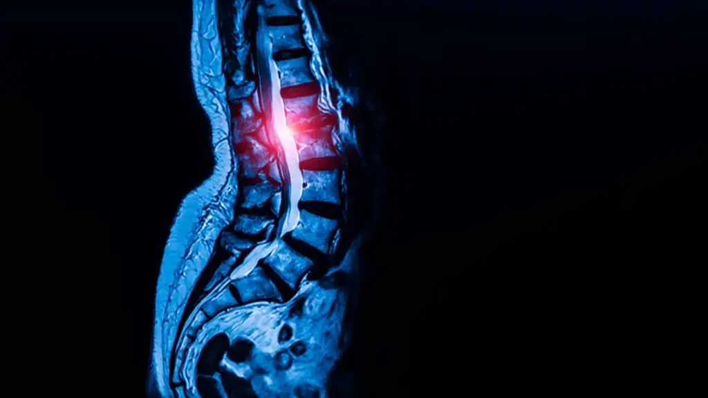
The debilitating condition affects an estimated 103 million people worldwide. It develops progressively when thickened ligaments reduce the available space inside the spinal canal. This dangerous narrowing puts chronic pressure on delicate nerves, frequently leading to severe pain, numbness, and significant difficulty walking.
The groundbreaking study, published in the journal Cell, fundamentally alters how scientists view connective tissue biology. More importantly, it raises the tantalizing possibility that directly targeting these newly discovered stem cells could eventually yield entirely new pharmacological treatments for spinal stenosis. One potential therapeutic approach under consideration involves existing medications, such as drugs already widely prescribed to control high blood pressure.
"While previous studies had proposed several candidate stem cells, none had definitively shown that a single cell population could both self-renew and generate the full spectrum of tendon and ligament cell types," said co-corresponding author Dr. Matthew Greenblatt, the Rohr Family Research Scholar, associate professor of pathology and laboratory medicine at Weill Cornell Medicine, and a pathologist at NewYork-Presbyterian/Weill Cornell Medical Center.
After successfully isolating and identifying the cells, the research team immediately investigated whether they might offer clinical clues to treating lumbar spinal stenosis. Currently, patients suffering from advanced stages of the condition have few viable medical options outside of invasive surgery once their nerve compression becomes severe.
"Identifying these specialized stem cells unlocks a new area of research that allows us to address this disease much more mechanistically, rather than just waiting until a patient’s condition worsens and requires surgery to relieve the nerve compression," said study co-corresponding author Dr. Sravisht Iyer, an associate professor of orthopedics at Weill Cornell Medicine and a spine surgeon at Hospital for Special Surgery. "The findings are exciting for their potential to change the way we deliver spinal care."
Finding the Stem Cells That Build Tendons and Ligaments
Dr. Greenblatt and his dedicated colleagues are no strangers to tracking down difficult-to-find skeletal cell populations. Back in 2018, the research group successfully identified the specific stem cell that initiates fracture repair within the outer layer of bone. They later followed that achievement by discovering the distinct stem cells involved in forming both the skull and the spine.
However, finding an equivalent progenitor cell for tendons and ligaments proved to be a significantly more formidable challenge. Unlike hard bone tissue, tendons and ligaments are packed with numerous fibroblast-like cells that look strikingly similar under a microscope, making it exceptionally difficult to distinguish one unique cell population from another.
"We analyzed thousands of individual cells and sorted them into individual cell types. Then we identified which one had the properties we associate with ‘stemness,’" Dr. Greenblatt explained, detailing the rigorous isolation process.
Within this context, "stemness" refers to the unique biological capacity of a rare cell population to continually renew itself over time while simultaneously producing the mature, specialized cells required to form and maintain healthy tendon and ligament tissue. Utilizing advanced models, the researchers discovered these elusive cells residing within a specialized anatomical region inside tendons and ligaments that appears to function as a natural reservoir for continuous tissue growth and repair.
Armed with insights gained from animal models, the research team then embarked on a search for equivalent cells within human tissues. Human ligament samples were carefully collected from tissue safely removed during surgical procedures by Dr. Iyer, utilizing specimens obtained exclusively from patients who had provided fully informed consent.
The researchers, which included first author Dr. Lingling Hu, a postdoctoral fellow operating within both Dr. Greenblatt’s and Dr. Iyer’s laboratories, ultimately confirmed that these human-derived cells possessed the same dual capabilities: they could successfully renew themselves and generate functional ligament cells.
Crucially, these specialized cells did not appear to be localized solely to the spinal region. Instead, they were found distributed broadly across the musculoskeletal system.
"We looked in the kneecap ligament; we looked at the Achilles tendon; and everywhere we looked, we found this cell," Dr. Greenblatt noted. "So, we think this is the universal stem cell for tendons and ligaments throughout the body."
How Stem Cells May Contribute to Spinal Stenosis
With the universal stem cell population identified, the researchers shifted their focus toward investigating whether these specific cells play a pathological role in the development of lumbar spinal stenosis.
To test this hypothesis, the team compared stem cells harvested directly from patients undergoing surgery for spinal stenosis against control cells collected from spinal ligaments removed from individuals who had suffered herniated discs but displayed no clinical signs or symptoms of stenosis. These patients also provided informed consent pre-operatively.
The analysis revealed a stark difference: ligaments harvested from individuals suffering from stenosis consistently contained a significantly higher concentration of the newly identified stem cells. Furthermore, when the researchers transplanted these stenosis-derived cells into experimental models, the cells produced an abundance of excess tendon cells compared to control stem cells taken from healthy individuals.
"Though spinal stenosis is a complex condition, this really showed us that these cells are contributing to the pathology," said Dr. Greenblatt, who is also an active member of the Sandra and Edward Meyer Cancer Center at Weill Cornell Medicine.
Seeking to understand the precise cellular mechanisms driving this overgrowth, the researchers peered deeper into the internal machinery of the cells. Stem cells associated with spinal stenosis exhibited measurably greater calcium signaling activity compared to their healthy counterparts.
Calcium signaling acts as a critical internal communication pathway that cells utilize to regulate core biological processes, including cellular growth, proliferation, and metabolic activity. To test causality, the scientists genetically manipulated healthy stem cells to artificially increase calcium signaling, successfully triggering excessive tissue growth that mimicked the pathological state.
A Possible Drug Target for Spinal Stenosis
Conversely, interrupting this pathway yielded promising results. When the researchers purposefully reduced calcium signaling within a mouse model of lumbar spinal stenosis, the abnormal, excessive cell growth was successfully blocked.
This pivotal finding strongly suggests that calcium signaling pathways could serve as a viable therapeutic target for pharmacological intervention. Furthermore, it raises the exciting possibility that calcium channel blockers—a well-established class of medications already widely prescribed across the globe to control high blood pressure—could one day be repurposed and formally studied as a non-surgical treatment for spinal stenosis. However, rigorous clinical trials will be fundamentally required before researchers can definitively determine whether this approach is both safe and effective for human patients.
"This is probably the first work that’s shown a potential therapeutic target for one of the most common spine conditions in the world," Dr. Iyer emphasized.
The clinical and scientific implications of the discovery may extend far beyond spinal stenosis alone. Moving forward, Dr. Greenblatt plans to investigate whether these exact same stem cells are implicated in a wider array of connective tissue disorders, including Marfan syndrome, a genetic condition that impacts connective tissues throughout the human body.
Additionally, the breakthrough could fundamentally help researchers decode why certain stubborn tendon and ligament injuries are notoriously difficult to heal naturally. Potential avenues for future scientific exploration include investigating chronic rotator cuff tears, severe Achilles tendon injuries, complex ligament reconstructions, and chronic tendon degeneration.
"Given that this cell appears to be the ultimate origin of all tendon and ligament cells, defects in this cell are likely at the heart of a wide range of tendon and ligament disorders," Dr. Greenblatt concluded.