



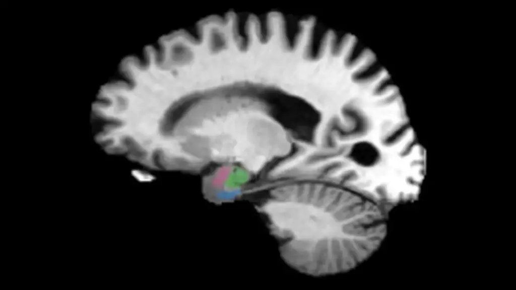
Alzheimer’s disease may begin leaving detectable, physical clues in the human brain as much as a decade before any memory problems or cognitive symptoms become apparent. In a significant step forward for neurodegenerative research, scientists at Johns Hopkins University have identified two specific brain regions that show clear signs of deterioration significantly earlier than the hippocampus, the area that has traditionally been the primary focus of medical examinations for evidence of the disease.
This discovery could eventually revolutionize how clinicians diagnose neurodegenerative conditions, allowing doctors to recognize Alzheimer’s years before clinical symptoms develop. By spotting the disease in its silent, early stages, future medical interventions could potentially begin at a time when treatments have the greatest chance of protecting the brain and preserving a patient’s cognitive health.
By examining hundreds of detailed brain scans, the research team found that the entorhinal cortex and the amygdala—two distinct regions heavily involved in processing memory and emotion—begin losing vital tissue much earlier than the hippocampus. These anatomical changes offer critical new insights into the natural progression of Alzheimer’s disease and point toward pathways for detecting the condition long before significant cognitive decline disrupts a patient’s daily life.
"We have found that there are early changes in the brain manifesting the clinical biomarkers of atrophy as much as a decade before there are clinical symptoms," said biomedical engineer and senior author Michael Miller. "It’s at least a 5-to-10-year play where your brain is changing. Imagine if we could start to treat the neurodegenerative changes at the 2, 3 or 4-year point rather than after 15 years when the changes are manifesting in memory symptoms. That’s the great promise here for your loved ones who are at risk."
The findings were recently published in the scientific journal NeuroImage Reports and are being showcased at Neuroscience 2026, the prestigious annual meeting of the Society for Neuroscience.
Detecting Alzheimer’s Before Memory Loss Begins
Alzheimer’s disease is widely recognized as the most common cause of dementia worldwide, affecting millions of aging individuals and their families. However, medical science has long understood that the pathology of the disease does not simply begin the moment an individual first experiences forgetfulness, confusion, or difficulty finding words. Instead, the cellular changes and protein abnormalities associated with Alzheimer’s can quietly accumulate and compound within the human brain for decades before any noticeable symptoms emerge on the surface.
Scientists increasingly believe that this extraordinarily long period of silent progression may represent a crucial, yet currently underutilized, window of opportunity for medical intervention. Therapeutics and lifestyle interventions that begin long before extensive, irreversible tissue damage occurs could theoretically do significantly more to preserve memory, executive function, and other vital cognitive abilities. However, taking practical advantage of that narrow window requires reliable, non-invasive ways to accurately identify the disease in its earliest, most manageable stages.
One of the most promising approaches in modern neurology involves the use of biomarkers—measurable biological indicators that can signal the presence, severity, or progression of an underlying disease process. In the context of Alzheimer’s research, these specific markers could eventually help clinicians reveal who is developing the condition years before symptoms become clinically apparent.
"Our goal is to be able to treat individuals before the symptoms of Alzheimer’s disease develop so that they may be delayed or even stopped," said Marilyn Albert, co-author of the study and director of the Johns Hopkins Alzheimer’s Disease Research Center.
Two Brain Regions Reveal Earlier Warning Signs
To investigate precisely where the earliest detectable structural changes occur within the central nervous system, the Johns Hopkins researchers analyzed hundreds of high-resolution magnetic resonance imaging (MRI) scans and positron emission tomography (PET) scans. These comprehensive datasets were collected through two major independent research initiatives: the Alzheimer’s Disease Neuroimaging Initiative and the Biomarkers of Cognitive Decline Among Normal Individuals study.
The extensive imaging archives tracked human subjects at various stages of health and disease progression over extended periods. The cohorts included individuals showing no cognitive symptoms whatsoever, patients experiencing mild cognitive impairment, and individuals who ultimately passed away from the disease, allowing researchers to build a longitudinal picture of how the brain changes over time.
Across both study groups, the researchers observed a striking and consistent pattern of degeneration. The entorhinal cortex and the amygdala, which are anatomically situated in close proximity to the hippocampus, demonstrated significantly greater relative tissue loss during the early phases of Alzheimer’s disease than the hippocampus itself.
This anatomical distinction is profoundly important for future diagnostic imaging protocols. While the hippocampus plays a central, well-documented role in memory formation and has traditionally served as the primary landmark when physicians search for Alzheimer’s-related brain damage, focusing exclusively on it may cause doctors to miss earlier warning signs.
The two newly highlighted regions perform critical, specialized functions of their own within the brain’s complex neural circuitry. The entorhinal cortex functions as a major hub that helps retrieve previously stored memories and acts as a gateway connecting the cerebral cortex with the hippocampus. Meanwhile, the amygdala contributes heavily to emotional processing and emotional memory, governing how powerful emotional experiences are interpreted, processed, and remembered over a lifetime.
The discovery that these structures undergo shrinkage earlier than the hippocampus suggests that clinicians and radiologists examining future brain scans could reveal signs of developing Alzheimer’s that might otherwise remain obscured when medical attention is focused solely on hippocampal volume.
Brain Shrinkage Linked to Alzheimer’s Tau Protein
Beyond simply cataloging the locations of early tissue loss, the researchers also uncovered a vital biological connection between this localized structural shrinkage and tau, a structural protein that has long been closely linked to the pathology of Alzheimer’s disease.
Under normal, healthy physiological conditions, tau proteins play a supportive role by helping to stabilize and maintain the internal microtubule structure of nerve cells, ensuring efficient nutrient transport and communication. In the brains of individuals with Alzheimer’s disease, however, the tau proteins undergo a chemical transformation, detaching from microtubules and abnormally accumulating into tangled clumps. These neurofibrillary tangles progressively interfere with healthy cellular function, ultimately leading to cell death.
Using advanced PET imaging techniques alongside digital pathology methods, the Johns Hopkins scientists discovered that the specific areas of gray matter shrinkage closely overlapped with the brain regions showing heavy tau-related protein changes. Gray matter is composed of a dense collection of nerve cell bodies, synapses, and dendrites, making it absolutely essential for processing information, directing physical actions, and managing cognitive tasks throughout the entire brain.
This clear, overlapping relationship significantly strengthens the scientific hypothesis that measuring structural changes in the entorhinal cortex and amygdala could serve as highly useful, early clinical indicators of developing Alzheimer’s disease.
The research team believes that quantifying tissue loss across both of these regions, rather than relying exclusively on traditional measurements of hippocampal atrophy, could eventually improve predictive models to determine which patients are most likely to experience cognitive decline as they age.
As modern neuroimaging technologies continue to evolve, becoming increasingly accessible, faster, and more cost-effective, this specialized diagnostic approach could become widely practical in everyday clinical settings. For individuals who carry a documented family history of Alzheimer’s disease or other recognized genetic and environmental risk factors, researchers envision a future in which medical professionals might routinely examine these specific brain structures as early as a person’s 30s or 40s.
"As biomarkers and treatments become increasingly available, knowing who is going to have cognitive decline is going to be really important," said co-author Meaghan Morris, a neuropathologist who specializes in the study of Alzheimer’s disease and related dementias. "This could really play into better risk stratification for identifying who is going to have cognitive decline in the near future — and treating people earlier and earlier to hopefully slow the course of the disease."
A Potential New Path Toward Earlier Alzheimer’s Treatment
Although the findings offer significant promise for the future of neurology and geriatric medicine, the researchers emphasize that substantial additional research is necessary before doctors can utilize these specific brain structural changes as a routine, foolproof diagnostic tool in standard clinical practice. The current study successfully identifies potential early biomarkers and structural warning signs, but it does not yet establish a fully realized clinical test capable of definitively predicting Alzheimer’s years in advance for an individual patient.
The logical next step for the research team is to investigate the biological mechanisms unfolding within these vulnerable brain regions at a much smaller, microscopic scale. The scientists want to gain a deeper understanding of the precise molecular changes surrounding abnormal tau accumulation and to map out how those molecular shifts directly drive the progressive, localized loss of vital brain tissue over time.
Uncovering the exact biological processes and cellular pathways involved could eventually reveal entirely new targets for pharmaceutical therapies designed specifically to interrupt the progression of Alzheimer’s disease long before extensive, permanent neural damage has a chance to occur.
"Our future work is to better show the molecular neighborhoods associated with these Tau protein changes as localized by these clinical biomarkers of atrophy," Miller said. "Once we know more about the molecular neighborhoods, we can develop therapies."