



You walk out of the grocery store, ready to head home and make dinner, carrying the routine mental checklist of an ordinary evening. But as you scan the crowded parking lot, a frustrating and increasingly familiar question comes to mind: Where did you leave your car? You stand by the automatic doors, trying to picture the exact spot, mentally retracing your steps through the aisles, but the memory stubbornly refuses to surface.
Remembering personal experiences like these depends heavily on episodic memory, a sophisticated cognitive faculty that allows humans to mentally time-travel and recall specific events from their lives. These memories form the foundational architecture of long-term memory, whether they involve a distant childhood milestone, such as the nervous first day of elementary school, or something that happened merely minutes earlier, like noting the row number where you parked your vehicle.
Yet, this vital cognitive ability notoriously tends to weaken as we grow older. The vivid sights, sounds, spatial cues, and emotional details associated with past experiences can become increasingly difficult to retrieve from the mind’s archives. For decades, scientists have grappled with the underlying mechanisms of this decline, and while some
degree of memory loss is widely recognized as a common, expected part of the natural aging process, these changes can sometimes accelerate and become far more severe in individuals developing Alzheimer’s disease.
Now, a compelling new study from researchers at the University of California, Berkeley, offers crucial new clues about what may be happening beneath the surface, deep inside the aging human brain. Neuroscientist Omer Sharon and his colleagues, working under the overarching direction of former Berkeley sleep researcher Matthew Walker, have discovered a tangible, measurable connection between disrupted brain waves during deep sleep, declining memory performance, and the subtle accumulation of tau, a notorious protein intricately associated with the pathology of Alzheimer’s disease.
The findings strongly suggest that the buildup of tau within the brain’s frontal cortex is directly linked to pathological changes in the coordinated electrical activity that normally works to preserve and lock in memories overnight.
"This is the first time we’ve shown this relationship between tau and how it messes with memory consolidation by attenuating traveling slow waves that originate in the frontal cortex," said Omer Sharon, a postdoctoral researcher at UC Berkeley’s Center for Human Sleep Science.
The groundbreaking research was published in the prestigious scientific journal Nature Neuroscience, opening up new avenues for understanding cognitive decline.
How Deep Sleep Helps the Brain Store Memories
Human sleep is far from a passive, uniform state of rest; rather, it involves several distinct, highly dynamic stages, each associated with vastly different patterns of electrical brain activity. Rapid eye movement, or REM, sleep is perhaps the most widely recognized stage, characterized by darting eyes beneath closed eyelids and the vivid dreaming that typically accompanies it. However, another crucial stage, known broadly as non-REM sleep, is exceptionally vital for maintaining healthy cognitive function and physiological restoration.
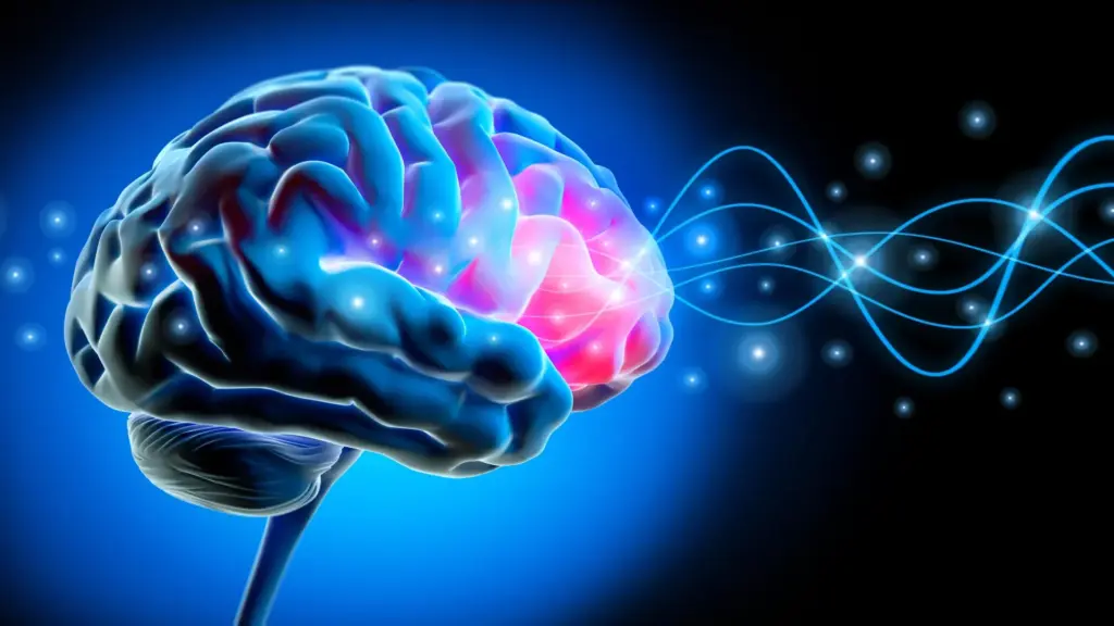
During this deeper stage of slow-wave sleep, when active dreaming is largely limited or entirely absent, massive populations of neurons briefly synchronize. They collectively become inactive and then fire back to life in a precisely timed, cascading sequence. These synchronized electrical shifts produce large, slow oscillations known as slow waves that ripple across the physical expanse of the brain, typically originating in the frontal cortex before sweeping outward.
"Each slow wave reflects an enormous population of neurons switching off and back on together," Sharon explained. "Our study shows that for memory, it’s critical that these events cascade in sequence across large parts of the brain. This coordinated shutdown has been observed only during deep sleep, and may be one reason why we sleep."
However, the researchers discovered that this carefully orchestrated neural activity becomes severely disrupted and less coordinated as tau protein progressively accumulates in the aging brain. Instead of traveling harmoniously together in broad, sweeping waves across vast regions of the cortex, the electrical waves become increasingly scattered, fragmented, and cover noticeably shorter distances. These structural changes in brain activity are strongly associated with weaker memory consolidation, declining everyday cognitive abilities, and the early stages of Alzheimer’s disease.
Alzheimer’s Protein Linked to Disrupted Brain Waves
To rigorously investigate this complex relationship, the team of scientists monitored the sleeping brain activity of human participants using high-density electroencephalograms, or EEGs, which record electrical patterns through the scalp. They carefully compared two distinct cohorts: cognitively healthy young adults in their early 20s and older participants ranging in age from their mid-60s to mid-70s.
The differences observed between the two age groups were striking. In the younger participants, robust groups of slow waves moved smoothly and synchronously across distances roughly equivalent to the width of a human hand on the scalp. Among the older adults, by contrast, the waves typically traveled significantly shorter distances and appeared far more independently, firing in a disjointed fashion rather than moving in synchronized, cooperative groups.
The research team also examined whether these structural alterations in sleep waves were directly associated with the accumulation of tau protein. Collaborating with Dr. William Jagust, a UC Berkeley professor emeritus of public health and neuroscience, the researchers utilized advanced positron emission tomography, or PET, scans to examine the cerebral architecture of the older participants. This sophisticated imaging technique introduces safe radioactive tracers into the bloodstream to reveal intricate biological activity and map the precise distribution of specific proteins within living human tissue.
The resulting scans revealed a clear pattern: greater tau buildup specifically within the frontal cortex was closely associated with a systemic breakdown in the longer, more expansive traveling waves normally observed during healthy deep sleep.
Crucially, these physiological changes were present even in people who had not developed clinical Alzheimer’s disease or experienced severe cognitive impairment.
"These people did not have Alzheimer’s," Sharon emphasized. "They had tau in their brains, but with subclinical impacts. Their memory decline was within the normal range for their age."
Sharon explained that early, Alzheimer’s-related biological changes can vary considerably in their severity across the population. The mere amount of tau present in the brain is tied to individual cognitive performance, even when a person’s daily memory abilities remain entirely within the expected, normative range for their chronological age.
Mysterious "Lonely Waves" Linked to Memory Loss
To move beyond structural brain imaging and test functional outcomes, the researchers wanted to determine whether these altered sleep patterns were directly associated with observable differences in how well people could actually retain and recall information.
To evaluate this, participants were given a series of learning tasks in the evening, memorizing pairs of associated words. The following day, after a full night of monitored sleep, they were tested on their ability to recall what they had learned, allowing the scientists to quantify how effectively their brains had consolidated the memories overnight.
The behavioral data matched the neurological findings: participants whose slow brain waves traveled shorter distances and appeared more isolated generally remembered significantly fewer word associations. The research team coined a descriptive term for these disconnected patterns, referring to them as "lonely waves."
Furthermore, the team tracked a subset of these participants over a longitudinal period spanning several years to observe how their brains changed over time. Among the individuals whose frontal cortex tau levels progressively increased during that window, the slow waves became noticeably less coordinated, and their inherent ability to retain newly learned information overnight steadily declined.
These comprehensive observations provide compelling empirical evidence of a robust relationship between tau accumulation, micro-structural changes in deep sleep brain activity, and worsening episodic memory. However, researchers caution that these findings do not yet establish definitively whether tau buildup directly causes memory loss, or whether the disrupted sleep waves themselves are the primary mechanism responsible.
Additional Tests Strengthen the Connection
Because PET scans are exceptionally costly, complex, and require specialized medical facilities and radioactive materials, the scientists sought an alternative methodological approach to investigate the relationship between Alzheimer’s-related proteins and sleep activity.
They established a collaboration with neurologist Yo-El Ju at Washington University in St. Louis to analyze cerebrospinal fluid drawn from an entirely separate group of older adults. The primary goal was to determine whether biochemical measurements of Alzheimer’s-associated proteins in spinal fluid would reveal patterns consistent with those observed via advanced brain imaging in California.
Unlike the high-resolution PET scans, the spinal fluid measurements could not isolate tau accumulation to specific anatomical regions like the frontal cortex. Nevertheless, the biochemical results pointed in the exact same direction.
Individuals whose sleeping brains produced a higher proportion of isolated slow waves exhibited significantly higher ratios of tau to amyloid—another pathological protein tightly linked to the development of Alzheimer’s disease—within their cerebrospinal fluid.
The striking consistency between these entirely independent cohorts and testing methods vastly strengthened the researchers’ confidence in the validity of their core conclusions.
"It’s collected in a totally different place and in a different cohort, so I think that adds to the strength of the evidence," Sharon noted.
Taken together, the overnight sleep recordings, neuroimaging brain scans, spinal fluid biochemical analyses, and rigorous memory assessments all converge on a single narrative: tau accumulation is intimately associated with a specific, measurable disruption of electrical brain activity during deep sleep.
"This longitudinal correlation suggests that Alzheimer’s disease pathology is associated with a disruption of this main sleep feature," Sharon said.
What Deep Sleep Reveals About Aging
Although everyday memory performance frequently declines as humans grow older, this new research indicates that the underlying biological narrative involves far more than simply the passage of time. Pathological changes long associated with the early development of Alzheimer’s disease—particularly the silent accumulation of tau protein in the cerebral regions responsible for generating widespread, synchronized slow waves—may help explain why some older adults experience significantly greater difficulty retaining memories than others.
"It’s not just about age," Sharon said. "It’s how much pathology you have in frontal areas where global waves originate."
Overall sleep quality also emerges as an essential part of the puzzle. Although sleep can often be notoriously difficult to define, quantify, or measure precisely in clinical settings, the physiological organization, trajectory, and physical movement of slow brain waves may ultimately provide critical diagnostic indicators of how effectively the human brain performs its essential nighttime memory functions.
The findings open the door to the possibility that monitoring changes in deep sleep electrical activity could one day help medical researchers better understand, detect, or identify early cognitive and memory problems before they fully manifest. They also point toward promising, novel targets for future therapeutic interventions aimed at protecting and preserving human memory as populations age.
For the time being, however, a central scientific question remains unanswered. Researchers must still determine whether accumulating tau directly contributes to the structural breakdown of synchronized sleep waves, whether structural changes in those waves actively influence the further accumulation of tau, or whether the relationship is driven by a broader, more complex underlying physiological process.
"We can see tau and lonely waves rising together over time," Sharon concluded. "What we cannot yet say is which leads—and that question will shape how we design interventions."
The study’s additional co-authors include Xi Chen, James Westphal, Chelsea Brown, and Vyoma D. Shah of UC Berkeley, along with Jason Dude of Washington University in St. Louis. Financial support for the research was provided by grants awarded by the National Institute on Aging at the National Institutes of Health, alongside fellowships from the Glenn Foundation for Medical Research and the Weill Neurohub.