



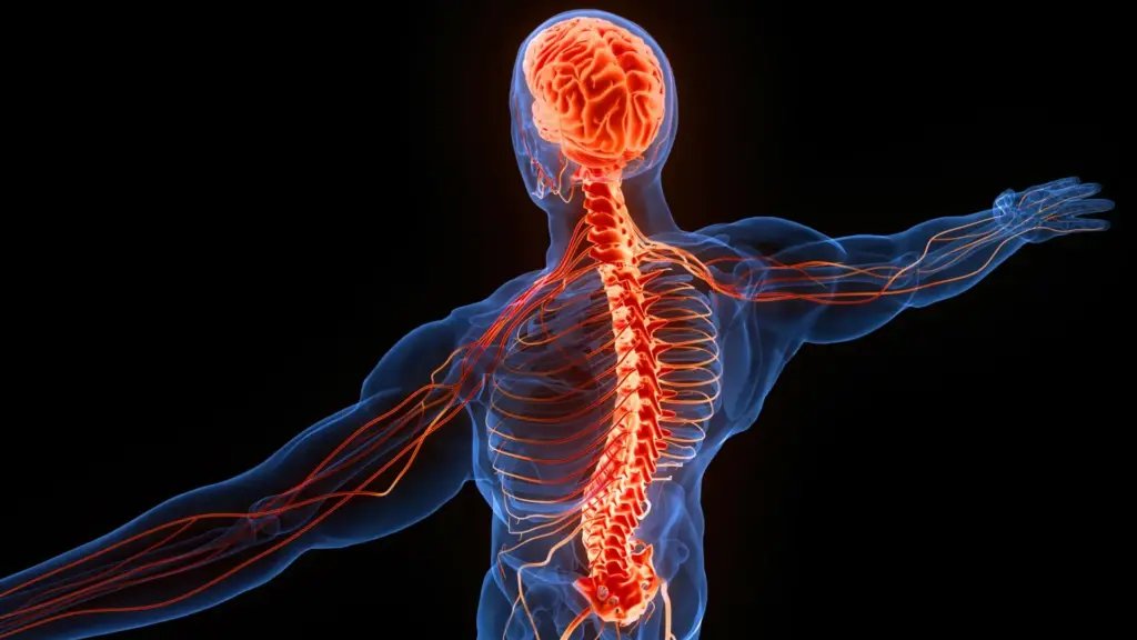
Why the left hemisphere of the human brain commands the right side of the body, and the right hemisphere rules the left, remains one of the most fascinating and enduring architectural puzzles of the nervous system. For centuries, this contralateral organization has baffled biologists, medical researchers, and philosophers alike. Now, a team of neuroscientists has proposed a groundbreaking explanation that stretches back hundreds of millions of years, tracing this peculiar cross-wired blueprint to the ancient visual systems of prehistoric aquatic creatures.
Dr. Martin Wessendorf, a neuroscientist and associate professor emeritus at the University of Minnesota, specializes in how the brainstem regulates neurons within the spinal cord. Alongside Daniel Wang, a specialist in cognitive neuroscience and neurodegenerative diseases, Wessendorf set out to examine the deep evolutionary origins of the brain’s crossed neural connections. Their findings, published in the journal Frontiers in Neuroanatomy, suggest that the human body’s cross-wired design may have originated simply because evolution repeatedly built new, more complex brain circuits around an already existing, highly ancient visual template.
The central nervous system—comprising the brain and spinal cord—is riddled with pathways that cross the midline. To understand why these contralateral connections exist, researchers must look deeply into the historical timeline of how sensory perception and motor control circuits evolved. The answer, according to the new study, may lie in the foundational mechanics of how primitive eyes captured light and how early vertebrate brains processed that visual data to ensure survival in a dangerous prehistoric world.
How the Brain Processes Visual Information
To trace the roots of this phenomenon, the researchers began by looking at the fundamental mechanics of vision. When light passes through the lens of an eye, the optical physics of the lens naturally project a reversed image onto the retina at the back of the eyeball. Consequently, objects appearing in our right visual field are projected onto the left side of the retina, while objects on our left are projected onto the right.
This visual data must then travel along the optic nerve toward an anatomical crossroads known as the optic chiasm. At this critical junction, nerve fibers are sorted so that information originating from the right visual field is successfully routed to the left side of the brain, while information from the left visual field is directed to the right hemisphere.
A portion of these vital optical signals eventually reaches an ancient midbrain structure called the optic tectum, known in mammals as the superior colliculus. This evolutionarily conserved region plays a crucial role in generating immediate, reflexive reactions to sudden visual stimuli, such as instantly noticing a predator or prey darting through the peripheral field of view.
Within the optic tectum, each side contains a precise neural map representing the opposite visual field. Furthermore, deeper layers within this same structure organize incoming physical sensations, known as somatosensory information, into corresponding maps that mirror the visual layout. Because the optic tectum is found across virtually every vertebrate species, science regards it as one of the oldest structures in the vertebrate nervous system. Its early emergence in evolutionary history could hold the primary key to understanding why countless other neural pathways across the body ultimately cross sides.
Why Sensory Signals Cross the Brain and Spinal Cord
Building upon this visual foundation, the researchers turned their attention to two major physiological pathways responsible for transmitting sensory data from the body up to the brain. Both of these vital pathways cross the center of the central nervous system, and both appear widely distributed across tetrapods—the broad classification of four-limbed animals that includes mammals, birds, reptiles, and amphibians.
The first of these is the dorsal column pathway, which is tasked with carrying sophisticated sensory information regarding gentle touch, physical vibration, and proprioception, the internal awareness of where different body parts are located in space. Signals gathered from the limbs and torso travel upward through the dorsal section of the spinal cord before making contact with another neuron situated in the brainstem, directly above the spinal cord. Only at this stage does the pathway finally cross over to the opposite side.
While fish lack this specific pathway entirely, scientists have successfully identified it in amphibians, reptiles, birds, and mammals. The consistent presence of the dorsal column pathway across such a diverse array of major animal lineages strongly indicates that this structural arrangement emerged very early during tetrapod evolution and has remained remarkably well-conserved over hundreds of millions of years.
The second major route, known as the spinothalamic tract, handles sensations that are critical for survival, including pain, itching, and specific mechanical touches. Unlike the dorsal column pathway, the spinothalamic tract crosses over to the opposite side within the spinal cord itself, rather than waiting to reach the brainstem. Once this crossing occurs, the sensory signals travel upward toward the thalamus, a central relay station positioned above the midbrain that coordinates sensory and motor information.
This particular crossed arrangement is readily observable in birds, mammals, and reptiles. Intriguingly, it also appears in certain cartilaginous fish, such as sharks and rays, indicating that elements of this sensory organization possess even deeper roots in evolutionary history than previously understood.
How the Brain Controls Movement Across the Body
The cross-wired nature of the nervous system is by no means limited to sensory perception. The researchers identified three major pathways dedicated to controlling skeletal muscles that also establish connections between one side of the brain and the opposite side of the spinal cord.
The first is the tectospinal pathway, which acts as a crucial conduit helping the body react swiftly to environmental cues. Because the optic tectum processes far more than just visual input, it also integrates sounds and physical sensations, arranging these diverse signals into maps that align seamlessly with its visual representation on the opposite side of the brain.
Consider the everyday scenario of preparing to cross a busy street when a car suddenly appears from the corner of your eye, prompting an instantaneous physical turn toward the vehicle. The tectospinal pathway is one of the primary neural circuits that drives this rapid defensive response, while also coordinating similar split-second reactions to unexpected sounds or physical touches. Movement signals originating deep within the optic tectum travel along nerve fibers directly to the opposite side of the spinal cord. This pathway is present in the majority of tetrapods as well as select cartilaginous fish.
Another critical circuit is the rubrospinal pathway, widely believed to assist animals in managing complex limb movements. It initiates within a midbrain structure known as the red nucleus and extends downward to the opposite side of the spinal cord. While most prominent in mammals, the rubrospinal pathway has also been documented in other tetrapods and certain cartilaginous fish.
The Evolution of Fine Motor Control in Humans
The third motor pathway examined by the researchers is the corticospinal tract, which holds extraordinary significance for humans because it underpins precise, intentional movements, including the delicate and intricate control of our fingers and hands. However, its evolutionary narrative is considerably more complex than those of its neurological counterparts.
All vertebrate animals possess a telencephalon, the forward region of the brain that accounts for the vast majority of overall brain volume in humans. Yet, only mammals possess a true cerebral cortex organized into six distinct cellular layers. This advanced six-layered cortex supports sophisticated cognitive tasks, such as synthesizing sensory data with stored memories, orchestrating complex movements, and processing language.
The corticospinal pathway projects outward from the cerebral cortex toward the spinal cord, though its functional role varies significantly across species. In the majority of mammals, the pathway primarily regulates sensory processing, with nerve fibers terminating in spinal cord regions that receive incoming bodily data, allowing the brain to filter and adjust sensations.
In primates—including humans—and select other animals capable of high-precision movements, the pathway also extends into regions of the spinal cord responsible for direct muscle activation. These evolutionary connections make intricate physical actions possible, such as the dexterous finger movements required for tool use and writing. Whether the corticospinal tract regulates sensory input or directly drives movement depending on the species, its fibers consistently cross the structural midline of the nervous system.
The researchers propose that humans may have unlocked advanced fine motor capabilities simply by repurposing an older neural circuit that originally evolved to manage sensory input. Rather than engineering an entirely new neurological system from scratch, evolutionary pressures appear to have assigned an existing pathway an innovative secondary purpose.
A 500-Million-Year-Old Explanation for the Brain’s Crossed Wiring
Taken together, these various sensory and motor pathways point toward a compelling and potentially surprising explanation for the brain’s unusual cross-wired design. The researchers hypothesize that the earliest visual circuits established an evolutionary precedent in which sensory and movement pathways were laid out across opposite sides of the nervous system. Subsequent neural circuits then inherited this structural template as they emerged and diversified, particularly within the tetrapod lineage.
Because every living vertebrate species possesses an optic chiasm and an optic tectum organized around data from the opposing visual field, the study’s authors suspect these anatomical structures were already fully established in primitive jawless fish during the Cambrian Period, roughly 538 to 486 million years ago. If this hypothesis proves correct, it means the foundational organization governing the brain’s crossed connections began taking shape more than half a billion years before modern humans ever walked the Earth.
Moreover, the hypothesis presents clear avenues for empirical testing. Scientists could theoretically examine fossilized neurocrania and skulls from ancient jawless fish for subtle anatomical evidence that might clarify whether these prehistoric animals already possessed an optic chiasm and a functioning optic tectum.
An Evolutionary Inheritance Rather Than a Functional Advantage?
This broad evolutionary perspective challenges a long-standing assumption in biology: the notion that every peculiar physiological trait must have evolved because it offered a direct, immediate survival advantage.
Evolution frequently modifies pre-existing anatomical structures rather than inventing entirely novel systems. Once ancient visual pathways established a cross-wired organizational standard, that established arrangement likely exerted a profound influence on how subsequent sensory and motor circuits developed. Specifically, the structural layout of the visual system—centered on the optic tectum—may have provided a reliable architectural scaffolding that later neurological pathways naturally followed.
Under this hypothesis, the brain’s crossed connections did not necessarily spread because controlling the opposite side of the body was inherently more efficient or effective. Instead, they may simply represent the lasting, inertial influence of an ancient template that evolution repeatedly utilized over vast stretches of geological time.
Ultimately, this ongoing scientific inquiry highlights one of the most remarkable examples of how the nervous system preserves its deep evolutionary history. The peculiar relationship linking the brain’s two hemispheres to the opposite sides of the body may trace its ultimate origins back to the primitive visual systems of aquatic organisms that swam the prehistoric oceans more than half a billion years ago.