
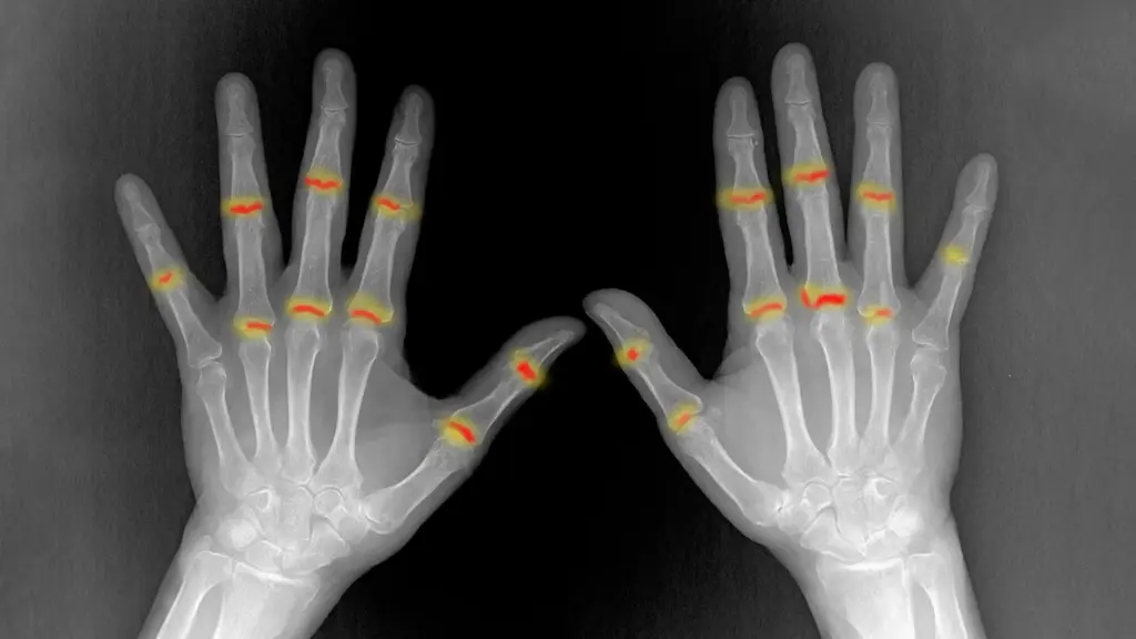
Researchers at the Kennedy Institute of Rheumatology have uncovered compelling evidence that certain human joints may carry a pre-programmed vulnerability to inflammatory arthritis long before a person is ever born. This groundbreaking discovery offers a potential solution to one of the most enduring medical mysteries surrounding rheumatoid arthritis: why the chronic autoimmune disease selectively targets specific joints while consistently leaving others completely untouched.
The study, titled "The embryonic origins of site-specific arthritis," was published in the prestigious journal Nature Immunology. Its findings strongly suggest that the answer to this medical enigma does not lie exclusively within the immune system, as previously assumed, but is also fundamentally rooted in the unique biological and structural differences built directly into individual joints during embryonic and fetal development.
Rheumatoid arthritis is a debilitating autoimmune condition in which the body’s immune system erroneously attacks the synovium, which is the specialized membrane and tissue lining the joints. This destructive autoimmune response produces severe pain, swelling, and chronic stiffness. Over extended periods, if left unchecked, the condition progressively damages vital cartilage, underlying bone, and the surrounding supportive tissues, leading to significant joint deformity and loss of mobility.
Some Joints Are Different Before Birth
To investigate why rheumatoid arthritis demonstrates such a distinct preference for particular skeletal locations, the research team focused their investigation on two specific types of finger joints that exhibit vastly contrasting patterns of disease susceptibility in adult patients.
The scientists closely compared proximal interphalangeal (PIP) joints, which are situated near the middle of the fingers and are frequently and severely affected by rheumatoid arthritis, with distal interphalangeal (DIP) joints located closer to the fingertips, which are typically spared from the ravages of the disease entirely.
Through meticulous analysis, the researchers discovered that the PIP joints possessed a noticeably larger volume of synovial tissue compared to their DIP counterparts. Furthermore, these vulnerable PIP joints contained a significantly higher concentration of PI16-positive (PI16+) fibroblasts, which represent a specialized subtype of connective tissue cells. Crucially, these distinct cellular and structural disparities were already fully present and observable well before birth. This foundational revelation strongly indicates that the local tissue environment established during a joint’s initial formation plays a critical role in determining whether destructive inflammation will be able to take hold and flourish decades later in life.
Christopher Buckley, Kennedy Professor of Translational Rheumatology at the University of Oxford, reflected on the significance of the findings, explaining that for decades, the medical community has recognized that rheumatoid arthritis selectively targets particular joints, yet one of the great unanswered questions has always been why this happens. He noted that their findings suggest the answer lies not only in the behavior of the immune system, but equally within the tissues themselves. The cellular and structural characteristics established during embryonic development help determine precisely where inflammation takes root later in life.
Mapping Developing Human Joints
To achieve this level of anatomical and cellular insight, the multidisciplinary research team deployed an array of cutting-edge technologies. They utilized single-cell sequencing, advanced image analysis software, and high-resolution 3D X-ray scanning to construct an extraordinarily detailed developmental map of human finger joints as they formed.
Examining joints during the active stages of development allowed the researchers to study entire joint structures in a level of minute detail that remains exceedingly difficult to achieve when working with fully mature adult tissue samples. During these early developmental windows, the team observed that developing joints consisted primarily of structural cells rather than the immune cells that typically dominate adult inflammatory sites. These structural building blocks included specialized fibroblasts and the progenitor cells directly responsible for forming healthy cartilage. The researchers then closely examined the intricate molecular signals that prompt these immature cells to differentiate into various specialized cell types.
One specific group of cells that drew intense scientific interest was the population of fibroblasts responsible for forming the synovial lining. Under normal physiological conditions, these specialized cells secrete essential lubricating substances that protect joints, minimize friction, and allow them to move smoothly and painlessly. However, in the context of autoimmune arthritis, these exact same protective cells undergo a pathological shift, beginning to behave abnormally and driving inflammation and tissue destruction.
Further biochemical and genetic analysis indicated that the synovial lining itself may actually develop from two distinct cellular sources: developing cartilage and the surrounding joint fibroblasts. Moreover, local microenvironmental conditions appeared to heavily influence this complex developmental process, including regions characterized by naturally occurring low oxygen levels, or hypoxia. Understanding these precise developmental signals could eventually provide crucial new clues regarding how synovial fibroblasts acquire their diverse functions, potentially revealing innovative therapeutic ways to reprogram and restore their normal, protective behavior in patients suffering from arthritis.
Specialized Cells in Arthritis Prone Joints
The researchers cataloged several critical differences between the arthritis-prone PIP joints and the disease-resistant DIP joints. Utilizing a specially developed digital image analysis tool, they demonstrated that PI16+ fibroblasts were markedly more abundant within PIP joints. These specialized cells were predominantly concentrated surrounding blood vessels and at critical anatomical locations where tendons and ligaments physically connect with nearby skeletal tissues.
Importantly, these PI16+ fibroblasts reacted differently to inflammatory biochemical signals compared to other fibroblast populations within the joint. Although both PI16+ fibroblasts and their PI16- counterparts shared a general pro-inflammatory response mechanism, the PI16+ cells exhibited distinct, unique changes in biological pathways directly involved in immune system regulation and the structural organization of tissue.
These structural and cellular divergences extended far beyond individual cell populations. By employing high-resolution 3D imaging capabilities at the Diamond Light Source facility located at the Harwell Science and Innovation Campus, the research team confirmed that PIP joints possessed a greater volume of synovial tissue, and that this tissue was architecturally organized in a fundamentally different manner from the synovium found in joints typically spared by rheumatoid arthritis.
When considered together, these profound differences spanning both distinct cell populations and overall tissue structure help provide a rational explanation for why chronic inflammation develops much more readily in certain joints than in others.
Dr. Sarah Davidson, a postdoctoral researcher at the Kennedy Institute and one of the primary authors of the study, emphasized that they found joints commonly targeted by rheumatoid arthritis already contain distinct cellular populations before birth. She noted that PI16+ fibroblasts were heavily enriched in vulnerable joints and reacted differently to inflammatory signals, meaning their specific anatomical location and cellular behavior heavily influence where the disease ultimately manifests.
A Developmental Clue to Rheumatoid Arthritis
Ultimately, these groundbreaking findings point toward a much broader conceptual framework for understanding why rheumatoid arthritis selectively targets particular parts of the human skeletal system. Rather than joint vulnerability being dictated entirely by systemic immune activity occurring later in life, an individual joint’s baseline tendency to develop chronic inflammation is also heavily dependent on the intrinsic cellular and structural features that were established years earlier while the joints were originally forming in the womb.
In essence, each individual joint constructs its own unique local biological environment during gestation, which subsequently helps dictate how susceptible that specific anatomical site will become to the destructive pressures of rheumatoid arthritis years or even decades into the future.
The extensive research project was led by dedicated scientists stationed at the Kennedy Institute of Rheumatology within the University of Oxford, working in close collaboration with academic colleagues from the University of Birmingham, University College London, and the Diamond Light Source. Financial support for the study was provided by the Medical Research Council.