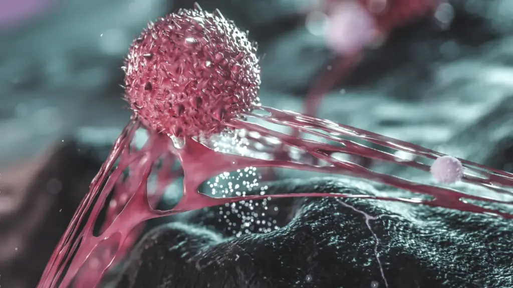
Researchers have constructed a groundbreaking, high-resolution cellular map of breast tumors that reveals a deeply complex microenvironment, featuring starkly divided regions filled with actively multiplying cancer cells alongside hidden pockets of dormant cells. Published in the journal Genome Medicine, the new findings demonstrate that these inactive cancer cells are frequently enveloped by specialized immune and connective tissue cells, creating localized microenvironments that may shield them from standard therapies.
The collaborative research project, spearheaded by scientists from the MRC Laboratory of Medical Sciences (LMS), Imperial College London, and the UCL Genetics Institute, highlights a critical challenge in modern oncology. Traditional cancer treatments are largely designed to hunt down and destroy fast-growing tumor cells. However, this new spatial mapping suggests that future therapies must evolve to simultaneously target dormant cancer cells and the local environments that allow them to persist. By doing so, medical researchers hope to not only achieve better immediate control over tumor growth but also significantly reduce the likelihood that the disease will return years down the line.
Breast tumors have long been recognized as complex, heterogeneous ecosystems composed of diverse cell types. Alongside the rapidly dividing cancer cells that drive tumor expansion, these masses contain immune cells, newly formed blood vessels, and a uniquely concerning subpopulation of cancer cells that remain unusually quiet.
These dormant, or quiescent, cells are known to possess the ability to survive aggressive treatment regimens. Once therapy concludes, these same cells can potentially reactivate, contributing to cancer recurrence or metastasis. To better understand this elusive population, researchers at the LMS, Imperial, and UCL set out to pinpoint the exact locations of these cells within untreated tumors, identify what distinguishes them from their proliferating counterparts, and map the specific cell types that tend to congregate around them.
Utilizing publicly available single-cell data, the interdisciplinary team successfully mapped out the architecture of breast cancer tumors. Their analysis uncovered distinct, isolated clusters of quiescent cells. Crucially, these clusters were not floating in isolation; rather, they were consistently surrounded by other cells that appear to function as a protective barrier, insulating the dormant cancer cells from external threats.
The danger posed by these hidden cells is a central focus for investigators in the field. Dr. Alexis Barr, co-lead author of the study and head of the Cell Cycle Control group at the LMS, emphasizes the unique threat these populations represent to patients undergoing therapy.
"Quiescent cancer cells are very dangerous," Dr. Barr explains. "These cells can hide from chemotherapy and then remain in this dormant quiescent state in the tumor, and then later reactivate to drive proliferation."
Under normal biological conditions, cancer cells can slip into this dormant state as a direct survival mechanism triggered by the harsh, stressful microenvironments inside a rapidly expanding tumor. As a tumor grows at an accelerated rate, the local blood supply and vital nutrient levels frequently fail to keep pace with the sheer demand of the multiplying mass. Faced with this metabolic crisis, certain cancer cells effectively put their cellular growth and division on hold, conserving their energy resources.
This biological strategy bears a striking resemblance to natural phenomena seen in the animal kingdom. Much like a bear hibernating through harsh winter conditions, these dormant cancer cells can hunker down and remain entirely inactive until external environmental conditions become more favorable. In the context of a patient, that window of opportunity often arrives after medical treatment has ended and the immediate selective pressure of chemotherapy has lifted.
Addressing this clinical hurdle is paramount for long-term patient survival, according to Dr. Barr. "If we want to achieve long-term control of peoples’ tumors and prevent tumor relapse, we have to focus on these dormant quiescent cancer cells, and have to understand more about them," she notes.
To investigate these hidden cell populations with unprecedented clarity, Dr. Barr partnered with Dr. Maria Secrier and her computational biology team at UCL. Together, the researchers constructed a comprehensive picture of the tumor landscape, examining both the cancer cells themselves and the surrounding web of immune and structural support cells.
To achieve this level of detail, the team combined single-cell RNA sequencing—a powerful technique that uncovers which specific genes individual cells are actively expressing—with spatial transcriptomics. This advanced spatial approach allows scientists to visualize the exact physical coordinates of cells within tissue architecture and identify which neighboring cells are in direct contact with them.
"We found cells that resemble therapy-resistant cells already residing in the tumor before we give any treatment," Dr. Secrier points out. This finding suggests that certain cellular characteristics associated with treatment resistance may already be hardwired into the tumor’s ecosystem before therapy even begins, rather than exclusively evolving as an adaptive response to the stress of drugs.
Intriguingly, the researchers observed this exact pattern across both highly aggressive forms of breast cancer and slower-developing classes of the disease. This was a particularly unexpected discovery, as cellular quiescence had historically been linked more closely with slower-growing tumors.
The scope of the analysis went far beyond the cancer cells, casting a wide net to examine the diverse supporting cell types that make up the broader tumor microenvironment. As the computational models mapped out the spatial relationships, a remarkably consistent pattern emerged across the samples.
Dormant cancer cells were frequently located in close physical proximity to CXCL10-positive macrophages—a specific subtype of immune cell—and myofibroblastic cancer-associated fibroblasts, which are known tumor-supportive structural cells. These surrounding cellular neighbors may have been recruited, reprogrammed, or chemically altered by the tumor in ways that directly protect the inactive cancer cells. One leading hypothesis is that these surrounding cells construct a robust physical or biological barrier, preventing cancer-killing immune cells or pharmacological treatments from successfully penetrating the niche to reach the dormant cells.
"The cancer cells are really encapsulated within these areas of macrophages and fibroblasts that we think act as shields for these dormant cancer cells," Dr. Secrier explains. However, she notes that the exact biological chain of events remains an open question for ongoing research. "We don’t yet know the direction of cause and effect: whether the surrounding cells push cancer cells into dormancy or if the cancer cells attract or alter their surroundings. It’s very likely coming from both sides."
These structural and behavioral differences point to a significant hurdle in current therapeutic approaches. The vast majority of standard chemotherapy drugs are designed to target and disrupt cellular division, meaning they work best against cells that are actively and rapidly multiplying. Dormant cells, by definition, bypass this vulnerability because they are not dividing, rendering them largely impervious to standard cytotoxic interventions.
Consequently, the findings strongly suggest that rapidly growing regions and dormant compartments within the very same tumor may require entirely different, tailored treatment strategies. Interestingly, the researchers also detected heightened activity within the complement pathway—an important component of the innate immune system—specifically localized inside these dormant cell niches. This biochemical clue raises the intriguing possibility that introducing targeted therapies designed to inhibit or modulate this pathway could make those previously sheltered areas vulnerable to intervention.
Similarly, the support cells clustered around the dormant cancer cells offer another promising avenue for drug development. However, before clinical applications can be designed, researchers must definitively determine whether these supporting cells are actively required to maintain cancer cell dormancy and how critical they are to the overall survival of the hidden cell populations.
"Different parts of the tumor will likely respond to different drugs," Dr. Secrier emphasizes. "If we understand what drug combinations we can use to target both the proliferative and the dormant areas, potentially that could be more successful than current therapies. This is giving us a first insight into how we can then intervene with different therapeutics that specifically target different areas of the tumor where the cells have adapted and have evolved differently."
Echoing this perspective, Dr. Barr underscores the need to rebalance research priorities in the field of oncology. "It is clearly important to focus on proliferative cancer cells, but we also need to understand this population of quiescent dormant cancer cells. And that’s been less studied."
While the theoretical frameworks and combination treatment ideas generated by this detailed computational analysis still require rigorous experimental testing in the laboratory, the implications are profound. By successfully locating treatment-resistant regions that pre-exist inside tumors and identifying the specific support cells that sustain them, scientists are laying the groundwork for a new generation of combination cancer therapies.
Ultimately, mapping out quiescent cells and the protective microenvironments that surround them brings researchers a step closer to designing precision treatments capable of simultaneously eradicating the rapidly proliferating portions of a tumor and neutralizing the stubborn, dormant cells that linger quietly behind, waiting for an opportunity to return.
The research was primarily funded by a UKRI Future Leaders Fellowship, the Medical Research Council, and the Biotechnology and Biological Sciences Research Council.